Selected News by :M.Hezarkhani MD.Urologist


21.June.2013

Cancer Res June 15, 2013 73; 3716

FGFR1 Is Essential for Prostate Cancer Progression and Metastasis

Feng Yang1, Yongyou Zhang3 Steven J. Ressler1, Michael M. Ittmann2, Gustavo E. Ayala2,

Truong D. Dang1, Fen Wang3, and David R. Rowley1

Authors' Affiliations: Departments of 1Molecular and Cellular Biology, 2Pathology and Immunology, Baylor College of Medicine, One Baylor Plaza; and 3Texas A&M University Health Science Center, Houston, Texas

+ Author Notes

Current address for G.E. Ayala: Department of Pathology, University of Texas Health Science Center, Houston, Texas; and Current address for Y. Zhang: Department of Hematology/Oncology, Case Western Reserve University, Cleveland, Ohio. 

The fibroblast growth factor receptor 1 (FGFR1) is ectopically expressed in prostate carcinoma cells, but its functional contributions are undefined. In this study, we report the evaluation of a tissue-specific conditional deletion mutant generated in an ARR2PBi(Pbsn)-Cre/TRAMP/fgfr1loxP/loxP transgenic mouse model of prostate cancer. Mice lacking fgfr1, in prostate cells developed smaller tumors that also included distinct cancer foci still expressing fgfr1 indicating focal escape from gene excision. Tumors with confirmed fgfr1 deletion exhibited increased foci of early, well-differentiated cancer and phyllodes-type tumors, and tumors that escaped fgfr1 deletion primarily exhibited a poorly differentiated phenotype. Consistent with these phenotypes, mice carrying the fgfr1 null allele survived significantly longer than those without fgfr1 deletion. Most interestingly, all metastases were primarily negative for the fgfr1 null allele, exhibited high FGFR1 expression, and a neuroendocrine phenotype regardless of fgfr1 status in the primary tumors.

Together, these results suggest a critical and permissive role of ectopic FGFR1 signaling in prostate tumorigenesis and particularly in mechanisms of metastasis.

©2013 American Association for Cancer Research.

 March 28, 2013; doi: 10.1158/0008-5472.CAN-12-3468

Cancer Res June 15, 2013 73; 3725  

Androgen Receptor-Independent Function of FoxA1 in Prostate Cancer Metastasis

Hong-Jian Jin1, Jonathan C. Zhao1, Irene Ogden1,Raymond C. Bergan1,2, and Jindan Yu1,2

Authors' Affiliations: 1Division of Hematology/Oncology, Department of Medicine, 2Robert H. Lurie Comprehensive Cancer Center, Northwestern University Feinberg School of Medicine, Chicago, Illinois  

FoxA1 (FOXA1) is a pioneering transcription factor of the androgen receptor (AR) that is indispensible for the lineage-specific gene expression of the prostate. To date, there have been conflicting reports on the role of FoxA1 in prostate cancer progression and prognosis. With recent discoveries of recurrent FoxA1 mutations in human prostate tumors, comprehensive understanding of FoxA1 function has become very important. Here, through genomic analysis, we reveal that FoxA1 regulates two distinct oncogenic processes via disparate mechanisms. FoxA1 induces cell growth requiring the AR pathway. On the other hand, FoxA1 inhibits cell motility and epithelial-to-mesenchymal transition (EMT) through AR-independent mechanism directly opposing the action of AR signaling. Using orthotopic mouse models, we further show that FoxA1 inhibits prostate tumor metastasis in vivo. Concordant with these contradictory effects on tumor progression, FoxA1 expression is slightly upregulated in localized prostate cancer wherein cell proliferation is the main feature, but is remarkably downregulated when the disease progresses to metastatic stage for which cell motility and EMT are essential. Importantly, recently identified FoxA1 mutants have drastically attenuated ability in suppressing cell motility.

Taken together, our findings illustrate an AR-independent function of FoxA1 as a metastasis inhibitor and provide a mechanism by which recurrent FoxA1 mutations contribute to prostate cancer progression.

©2013 American Association for Cancer 

 April 30, 2013; doi: 10.1158/0008-5472.CAN-12-4563

Cancer Res June 15, 2013 73; 3604

BMP-6 in Renal Cell Carcinoma Promotes Tumor Proliferation through IL-10–Dependent M2 Polarization of Tumor-Associated Macrophages

Jae-Ho Lee1, Geun Taek Lee1, Seung Hyo Woo1, Yun-Sok Ha1,2, Seok Joo Kwon1, Wun-Jae Kim2, and Isaac Yi Kim1

Authors' Affiliations: 1Section of Urologic Oncology, The Cancer Institute of New Jersey and Robert Wood Johnson Medical School, New Brunswick, New Jersey; and 2Department of Urology, Chungbuk National University College of Medicine, Cheongju, Korea

Dysregulated bone morphogenetic proteins (BMP) may contribute to the development and progression of renal cell carcinoma (RCC). Herein, we report that BMP-6 promotes the growth of RCC by interleukin (IL)-10–mediated M2 polarization of tumor-associated macrophages (TAM). BMP-6–mediated IL-10 expression in macrophages required Smad5 and STAT3. In human RCC specimens, the three-marker signature BMP-6/IL-10/CD68 was associated with a poor prognosis. Furthermore, patients with elevated IL-10 serum levels had worse outcome after surgery.

Together, our results suggest that BMP-6/macrophage/IL-10 regulates M2 polarization of TAMs in RCC.

©2013 American Association for Cancer Research.


Endothelial Cells Enhance Prostate Cancer Metastasis via IL-6→Androgen Receptor→TGF-β→MMP-9 Signals

Xiaohai Wang1,2, Soo Ok Lee1, Shujie Xia2, Qi Jiang1,2, Jie Luo1, Lei Li1, Shuyuan Yeh1, and Chawnshang Chang1,3 

Authors' Affiliations: 1George Whipple Lab for Cancer Research, Departments of Pathology, Urology, and Radiation Oncology, and The Wilmot Cancer Center, University of Rochester Medical Center, Rochester, New York; 2Department of Urology, Shanghai First People's Hospital, Shanghai Jiaotong University, Shanghai, China; and 3Sex Hormone Research Center, China Medical University/Hospital, Taichung, Taiwan  

Although the potential roles of endothelial cells in the microvascules of prostate cancer during angiogenesis have been documented, their direct impacts on the prostate cancer metastasis remain unclear. We found that the CD31-positive and CD34-positive endothelial cells are increased in prostate cancer compared with the normal tissues and that these endothelial cells were decreased upon castration, gradually recovered with time, and increased after prostate cancer progressed into the castration-resistant stage, suggesting a potential linkage of these endothelial cells with androgen deprivation therapy. The in vitro invasion assays showed that the coculture of endothelial cells with prostate cancer cells significantly enhanced the invasion ability of the prostate cancer cells. Mechanism dissection found that coculture of prostate cancer cells with endothelial cells led to increased interleukin (IL)-6 secretion from endothelial cells, which may result in downregulation of androgen receptor (AR) signaling in prostate cancer cells and then the activation of TGF-β/matrix metalloproteinase-9 (MMP-9) signaling. The consequences of the IL-6→AR→TGFβ→MMP-9 signaling pathway might then trigger the increased invasion of prostate cancer cells. Blocking the IL-6→AR→TGFβ→MMP-9 signaling pathway either by IL-6 antibody, AR-siRNA, or TGF-β1 inhibitor all interrupted the ability of endothelial cells to influence prostate cancer invasion.

These results, for the first time, revealed the important roles of endothelial cells within the prostate cancer microenvironment to promote the prostate cancer metastasis and provide new potential targets of IL-6→AR→TGFβ→MMP-9 signals to battle the prostate cancer metastasis.

©2013 American Association for Cancer Research.
Diabetes Mellitus and Prostate Cancer Risk; A Nationwide Case–Control Study PCBaSe Sweden

NEWS/Cancer Epidemiol Biomarkers Prev June 2013 22; 1102

Katja Fall1,2, Hans Garmo3, Soffia Gudbjörnsdottir6, Pär Stattin7, and Björn Zethelius4,5 

1Clinical Epidemiology and Biostatistics, Örebro University Hospital and University, Örebro, Sweden; 2Harvard School of Public Health, Boston, Massachusetts; 3Regional Oncology Center; 4Department of Public Health and Caring Sciences/Geriatrics, Uppsala University; 5Medical Products Agency, Uppsala; 6Department of Medicine, Sahlgrenska University Hospital, Gothenburgh University, Göteborg, Sweden; and 7Department of Surgery, Urology Service, Memorial Sloan-Kettering Cancer Center, New York City, New York  

Background: Diabetes mellitus (DM) increases the risk for cancer at almost all sites, but data on the association with prostate cancer are inconsistent.

Methods: We assessed the risk of a prostate cancer diagnosis among men with type 2 (T2)DM in a nationwide population-based case-control study including 44,352 men with prostate cancer identified through the Prostate Cancer data Base Sweden (PCBaSe) between 2002 and 2006 and 221,495 age-matched men from the general population.

Results: Overall, the risk of prostate cancer among men with T2DM was lower than among men without T2DM [OR, 0.80; 95% confidence interval (CI), 0.76–0.85]. The risk decreased with longer disease duration and was observed across all tumor risk categories, although most clearly among men with low risk tumors (OR, 0.71; 95% CI, 0.64–0.80). The risk for prostate cancer was reduced among diabetic men on dietary treatment only (OR, 0.89; 95% CI, 0.80–0.99) but more markedly among men on oral hypoglycemic agents (OR, 0.80; 95% CI, 0.74–0.87) and insulin (OR, 0.72; 95% CI, 0.69–0.81). Obese diabetic men (BMI > 30 kg/m2) showed a reduced risk (OR, 0.72; 95% CI, 0.65–0.80) compared with men without diabetes. There was a trend of decreasing risk with increasing levels of HbA1c (P < 0.05).

Conclusions: This nationwide study confirmed a reduced risk of being diagnosed with prostate cancer among men with T2DM, especially for low-risk tumors. An altered hormonal milieu is a plausible explanation, although the possibility of decreased prostate cancer detection among diabetic men cannot be ruled out.

©2013 American Association for Cancer Research.

Selective estrogen receptor modulators and BPH

Edited by:Mohammad  Hezarkhani  MD,Urologist

Board-Certified of Urology,Tehran  University ,The Member  of  Iranian  Urological  Association

Madaen Hospital  Tehran Iran

Mohammad.hezarkhani@yahoo.com

www.facebook.com/mohammad.hezarkhani.1

June,20 , 2013

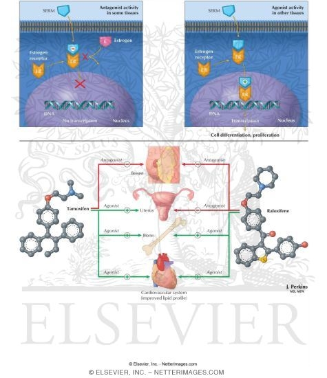
Selective estrogen receptor modulators (SERMs) are a class of compounds that act on the estrogen receptor. A characteristic that distinguishes these substances from pure receptor agonists and antagonists is that their action is different in various tissues, thereby granting the possibility to selectively inhibit or stimulate estrogen-like action in various tissues. Phytoserms are SERMs from a botanical source.

Estrogenic compounds span a spectrum of activity ranging from:

  • full agonists (agonistic in all tissues) such as the natural endogenous hormone estrogen
  • mixed agonists/antagonistics (agonistic in some tissues while antagonist in others) such as tamoxifen (a SERM)
  • pure antagonists (antagonistic in all tissues) such as fulvestrant (ICI-182780).

The mechanism of mixed agonism/antagonism may differ depending on the chemical structure of the SERM, but, for at least for some SERMs, it appears to be related to (1) the ratio of co-activator to co-repressor proteins in different cell types and (2) the conformation of the estrogen receptor induced by drug binding, which in turn determines how strongly the drug/receptor complex recruits co-activators (resulting in an agonist response) relative to co-repressors (resulting in antagonism).

 For example, the prototypical SERM tamoxifen acts as an antagonist in breast and conversely an agonist in uterus. The concentration of steroid receptor co-activator 1 (SRC-1; NCOA1) is higher in uterus than in breast, therefore SERMs such as tamoxifen are more agonistic in uterus than in breast. In contrast, raloxifene behaves as an antagonist in both tissues. It appears that raloxifene more strongly recruits co-repressor proteins and consequently is still an antagonist in the uterus despite the higher concentration of co-activators relative to co-repressors.

The actions of SERMs on various tissues:

  • Bone turnover and postmenopausal osteoporosis respond favorably to most SERMs, although premenopausal women may experience bone loss with some SERMs including tamoxifen.
  • Breast - all SERMs decrease breast cancer risk, and tamoxifen is mainly used for its ability to inhibit growth in estrogen receptor-positive breast cancer.
  • Cholesterol and triglycerides - levels respond favorably to SERMs.
  • Deep venous thrombosis - the risk may be elevated in at least some SERMs.
  • Hot flashes are increased by some SERMs.
  • Pituitary gland - clomifene blocks estrogen action, leading to an increase of follicle-stimulating hormone and luteinizing hormone.
  • Uterus - tamoxifen may increase endometrial carcinoma risk, but raloxifene and femarelle do not. Data on toremifene and clomifene is insufficient.
  • There is ample evidence in literature that suggests the role of estrogens in BPH development and management through the different tissue and cell-specific receptors. This article reviews beneficial actions of selective estrogen receptor modulator (SERM) and ERβ-selective ligands, which have been demonstrated through in vitro studies using human prostate cell lines and in vivo animal studies. SERMs have anti-proliferative, anti-inflammatory and pro-apoptotic mechanisms in BPH, and also act by inhibiting various growth factors, and thus represent a unique and novel approach in BPH management directed at estrogen receptors or estrogen metabolism.

References:

1- Prostate Cancer and Prostatic Diseases , (18 June 2013) | doi:10.1038/pcan.2013.17

Selective estrogen receptor modulators for BPH: new factors on the ground/M Garg, D Dalela, D Dalela, A Goel, M Kumar, G Gupta and S N Sankhwar

2-Wikipedia.The Free Encyclopedia, Selective estrogen-receptor modulator/27.Feb.2013

Exposure to Bisphenol A Increases Risk of Prostate Cancer

Edited by:Mohammad  Hezarkhani  MD,Urologist

Board-Certified of Urology,Tehran  University ,The Member  of  Iranian  Urological  Association

Madaen Hospital  Tehran Iran

Mohammad.hezarkhani@yahoo.com

www.facebook.com/mohammad.hezarkhani.1

June,20 , 2013


Bisphenol A (BPA) is a man-made carbon-based synthetic compound with the chemical formula (CH3)2C(C6H4OH)2 belonging to the group of diphenylmethane derivatives and bisphenols.BPA is used to make certain plastics and epoxy resins.  

BPA-based plastic is clear and tough, and is used to make a variety of common consumer goods (such as baby and water bottles, sports equipment, and CDs and DVDs) and for industrial purposes, like lining water pipes. Epoxy resins containing BPA are used as coatings on the inside of many food and beverage cans. It is also used in making thermal paper such as that used in sales receipts. 

It is part of the bisphenols group of chemical compounds with two hydroxyphenyl functionalities. It is a colorless solid that is soluble in organic solvents, but poorly soluble in water. Bisphenol A has a vapor pressure of 5×10⁻⁶ Pa. BPA exhibits hormone-like properties that raise concern about its suitability in consumer products and food containers

Bisphenol A is used primarily to make plastics, and products using bisphenol A-based plastics have been in commercial use since 1957. At least 3.6 million tonnes (8 billion pounds) of BPA are used by manufacturers yearly. It is a key monomer in production of epoxy resinsand in the most common form of polycarbonate plastic. Bisphenol A and phosgene react to give polycarbonate under biphasic conditions; the hydrochloric acid is scavenged with aqueous base: 

Diphenyl carbonate may be used in place of phosgene. Phenol is eliminated instead of hydrochloric acid. This transesterification process avoids the toxicity and handling of phosgene.

Polycarbonate plastic, which is clear and nearly shatter-proof, is used to make a variety of common products including baby and water bottles, sports equipment, medical and dental devices, dental fillings sealants, CDs and DVDs, household electronics, eyeglass lenses, foundry castings and for lining water pipes.

 BPA is also used in the synthesis of polysulfones and polyether ketones, as an antioxidant in some plasticizers, and as a polymerization inhibitor in PVC. Epoxy resins containing bisphenol A are used as coatings on the inside of almost all food and beverage cans; however, due to BPA health concerns, in Japan epoxy coating was mostly replaced by PET film. Bisphenol A is also a precursor to the flame retardant tetrabromobisphenol A, and formerly was used as a fungicide.

Bisphenol A is a preferred color developer in carbonless copy paper and thermal paper, with the most common public exposure coming from some thermal point of sale receipt paper. When used in thermal paper, BPA is present as “free” (i.e., discrete, non-polymerized) BPA, which is likely to be more available for exposure than BPA polymerized into a resin or plastic. Upon handling, BPA in thermal paper can be transferred to skin, and there is some concern that residues on hands could be ingested through incidental hand-to-mouth contact.  

Furthermore, some studies suggest that dermal absorption may contribute some small fraction to the overall human exposure. European data indicate that the use of BPA in paper may also contribute to the presence of BPA in the stream of recycled paper and in landfills.  

It has been suggested that the environmental factor BPA may play an important role in the initiation (Ho et al. [2006]) and progression of PCa and in hormonal therapy bypass (Wetherill et al. [2005]). At the level of PCa cells, BPA was able to induce androgen-independent tumor cell proliferation and reduced therapeutic efficacy in xenograft models (Wetherill et al. [2006]). While these data point toward the potential for BPA to assist tumor cells in escaping therapy, the molecular mechanisms of this process were not well-known.

This report demonstrates for the first time that the environmentally relevant concentrations of BPA (1–10 nM) induce cell migration by modulating the cell calcium signalling. These data highlight the previously unrecognized action of BPA in the progression of human PCa, thereby providing strong support for the growing recognition of the adverse effects of BPA on human health.

Invasion and metastasis are major events underlying cancer morbidity and mortality (Molloy and Van ’t [Veer 2008]). Because of the widespread metastasis in advanced cancer patients, where a resistance is observed to conventional therapies, the mortality rate remains extremely high and warrants new strategies to intervene in the metastatic cascade.

Thus, an enhanced understanding of the molecular events in the pathogenesis of PCa will offer improved diagnosis, prognosis, therapy and prevention measures of the disease, that will ultimately help us to eliminate PCa metastasis. A common regulatory point in several signal transduction pathways is intracellular calcium homeostasis. One approach could be to focus on the intracellular signalling pathways underlying the metastatic process. Several data have clearly shown the involvement of calcium entry in cancer and non-cancer cell migration (Bisaillon et al. [2010]; Li et al. [2011]; Schaff et al. [2010]; Yang et al. [2009]).

In the present work, we present conclusive evidence for the first time that the pre-treatment of human PCa cells with environmentally relevant concentrations of BPA (1–10 nM) induces their migration .By calcium imaging technique, we show that BPA pre-treatment induces an amplification of Store-Operated Calcium Entry (SOCE) in LNCaP cells .RT-PCR and Western blot experiments allowed us to identify those ion channel proteins up-regulated by BPA pre-treatments.

These channels include Orai1, a protein known to constitute an important actor in SOCE in various cell systems including human PCa cells .Meanwhile, in our studies, we failed to observe any direct effect of BPA on the rate of basal calcium whereas in other cell systems, BPA or its derivatives induced a calcium increase.

 In TM4 Sertoli cells, a direct application of a derivative compound of BPA, Tetrabromobisphenol A (TBBPA), a commonly used brominated flame retardant (BFR), induced an increase in the basal free calcium rate originating from internal stores (Ogunbayo et al. [2008]).

In pituitary tumor cells (GH3/B6/F10 rat Somatomammotropes), Kochukov et al. ([2009]) showed that BPA at a 1 nM concentration induced a great increase in Ca2+ oscillation frequency, the activation of MAPK pathways (ERK1/2) and subsequently a PRL release (Kochukov et al. [2009]). Similar results were reported by Bulayeva et al. ([2005]) and Wozniak et al. ([2005]) where the authors demonstrated that the BPA-induced Ca2+ influx was strictly dependent on membrane Estrogen Receptor (mER-α) and mediated by L-type voltage-gated Ca2+ channels in pancreatic β cells (Bulayeva et al. [2005]; Wozniak et al. [2005]).

The LNCaP cells used in our present work do not express L-type voltage-gated Ca2+channels (non-excitable cells). This is probably the reason why a direct application of BPA on LNCaP and LNCaP C4.2 cells failed to induce a direct calcium response.

In the present work, when the human PCa cells were pre-treated with BPA (1–10 nM), an increase in SOCE and a remodelling of ion channel expression was observed. The alterations of the ion channel expression could be mediated by a stimulation of a signal transduction pathways leading to the activation of nuclear transcription factors. Published data suggest several transduction pathways activated by BPA.

In PCa cells, BPA has been shown to be an agonist for mutant androgen receptor (AR-T877A) expressed in recurrent PCa (Wetherill et al. [2002,,2005,,2006]), and in the LNCaP cell line used in our studies. According to the authors, BPA induces cell proliferation in cells expressing the mutated AR. The clinical ramifications of BPA activating tumor-derived mutant ARs and inducing androgen-independent tumor cell proliferation may be substantial, as BPA can reduce therapeutic efficacy in xenograft models (Wetherill et al. [2006]).

In our experiments, the DHT induced the expression of Orai1 and BPA appears to mimic these effects on the canonical AR ligand (DHT). However, all the effects of BPA could not be mediated by the activation of AR. In a recent work, Hess-Wilson et al. ([2007]) showed clearly that BPA and DHT elicited distinct transcriptional signatures in PCa cells expressing the BPA-responsive mutant AR-T877A, even if some common genes were activated by both DHT and BPA in LNCaP cells (Hess-Wilson et al. [2007]).

These observations could explain the cell migration and Orai1 expression induced by BPA in androgen-independent human PCa cells PC-3, where the AR is absent. BPA could thus activate other signal transduction pathways than the AR activation, to induce the effects observed in our studies on androgen-insensitive PCa cells.

The study of the involvement of other transduction pathways than those involving the AR receptor (growth factor signalling pathways, …) in the effects of BPA in androgen-dependent and androgen-independent cells needs further extensive investigations in the future. In this context, BPA was reported to induce the phosphorylation of extracellular signal–regulated kinase (ERK), c-Jun N-terminal kinase (JNK), and nuclear translocation of the nuclear factor (NF)-κB, in mouse hippocampal HT-22 cells (Lee et al. [2008]).

 Functional NF-κB-binding sites in promoter regions of STIM1 and Orai1 have been identified and the expression of the Orai1 calcium channel was reported to be positively modulated by NF-κB (Eylenstein et al. [2012]). Subsequently, the store-operated Ca2+ entry was similarly increased by overexpression of p65/p50 or p65/p52, and decreased by treatment with the NF-κB inhibitor, Wogonin. BPA could thus interfere with the growth factors' signal transduction to activate the PI3K/AkT pathway and thereby induce the activation of the NF-kB transcription factor, leading in turn to an up-regulation of the expression of ion channels including Orai1.

 In this context, authors have shown that the activation of mER-α induces the activation of the PI3K/Akt signalling cascade (Simoncini et al. [2003]; Stirone et al. [2005]) and BPA is shown to activate mER-α (Bouskine et al. [2009]; Quesada et al. [2002]). The activation of the mER-α in androgen-dependent (LNCaP) and androgen-independent (PC-3) PCa cells may thus induce the PI3K/Akt signalling cascade which leads to the activation of the NF-kB transcription factor and Orai1 gene expression. Several works have also demonstrated the stimulation of the PKA/CREB pathways by nanomolar concentrations of BPA through the activation of mER-α (Bouskine et al. [2009]; Quesada et al. [2002]).

However, the involvement of this pathway in the expression of ion channels needs further investigation.  BPA pre-treatment induced an increase in BKCa and IKCa1 Ca2+-activated potassium channel expression in LNCaP cells. We previously showed that the IKCa1 Ca2+-activated potassium channels are involved in SOCE in LNCaP and PC-3 human PCa cells (Lallet-Daher et al. [2009]).

These potassium channels could constitute a functional complex with Orai1 protein to promote calcium entry and cell migration. A study is in progress in our lab to show the functional up-regulation and involvement of these Ca2+-activated potassium channels (IKCa1 and BKCa) in cell migration in the BPA-treated LNCaP and PC-3 cancer cells.

Receptor-mediated activation of phospholipases C by the factors present in the serum leads to IP3-mediated depletion of Ca2+ from the ER, which in turn stimulates Ca2+ influx through the plasma membrane involving Orai1/STIM1 complex formation (Varnai et al. [2009]). Recent works have elegantly demonstrated that STIM1, Orai1, and SOCE play critical roles in the migration of a number of cell types in cancer and non-cancer cells (Bisaillon et al. [2010]; Li et al. [2011]). These observations support our data where BPA, by up-regulating the ion channels expression increases the SOCE developed by PCa cells in response to factors present in serum.

 BPA-induced cell migration is dependent on the calcium entry and the use of pharmacological tools suggests the involvement of SOCE channels in the effects of BPA on cell migration .Increase in cytoplasmic calcium induced by BPA may have several types of impacts which trigger cell migration, including the induction of the up-regulation of their gene and protein expression, secretion and the activation of the enzymes such as metalloproteinases (MMP2, MMP9), which are involved in cell migration.

 The MMP proteins are clearly shown to be dependent on calcium for their expression (calcium/Calcineurin/NFAT, …), their processing and their activity (Collier et al. [1988]; Mukhopadhyay et al. [2004]; Stetler-Stevenson et al. [1989]). These observations suggest that the increase in calcium entry induced by BPA pre-treatment could promote all these processes leading to cell migration.

For the first time,researcher demonstrate the expression of Orai1 proteins in human PCa tissues .As shown, a strong immuno-staining of Orai1 protein was found in epithelial cells of the acini and also in the stromal cells. Thus, stromal cells could also be influenced by BPA impregnation. Given the importance of the epithelium-stroma (reactive stroma) in the progression of cancer, the potential effects of BPA on calcium signalling and on the secretion of growth factors by these cells need further investigation.

Researcher believe that BPA actually reprograms the stem cells to be more sensitive to estrogen throughout life, leading to a life-long increased susceptibility for diseases including cancer.

References:

1- cancer Bisphenol A stimulates human prostate cell migration via remodelling of calcium signalling

Sandra Derouiche1,2, Marine Warnier1,2, Pascal Mariot1,2, Pierre Gosset3, Brigitte Mauroy4, Jean-Louis Bonnal4, Christian Slomianny1,2, Philippe Delcourt1,2, Natalia Prevarskaya1,2 and Morad Roudbaraki1,2* 1 Inserm, U-1003, Equipe labellisée par la Ligue Nationale contre le cancer, Villeneuve d’Ascq, France ,2 Laboratory of Excellence, Ion Channels Science and Therapeutics; Université Lille I Sciences et Technologies, Villeneuve d’Ascq, France ,3 Laboratoire d’Anatomie et de Cytologie Pathologique du groupement hospitalier de l’Institut Catholique de Lille, Faculté Libre de Médecine, Lille, France ,4 Service d’Urologie de l’hôpital St-Philibert, Lomme, France /SpringerPlus 2013, 2:54 doi:10.1186/2193-1801-2-54

2-Wikipedia, The Free Encyclopedia,Bisphenol A   18/June/2013

3- Exposure to BPA in Developing Prostate Increases Risk of Later Cancer: Ubiquitous Plasticizers May      Have Long-Term Health Effects/ Prins, professor of physiology and director of the andrology laboratory in urology at the UIC College of Medicine.17/June/2013