LATEST MEDICAL NEWS

 
LATEST MEDICAL NEWS
 
12.DECEMBER
 

The immune cells that help tumors instead of destroying them

Lung cancer is the leading cause of cancer-associated deaths. One of the most promising ways to treat it is by immunotherapy, a strategy that turns the patient's immune system against the tumor. In the past twenty years, ...

2 hours ago
popularity comments

Discovery deepens understanding of brain's sensory circuitry

Because they provide an exemplary physiological model of how the mammalian brain receives sensory information, neural structures called "mouse whisker barrels" have been the subject of study by neuroscientists around the ...

2 hours ago
popularity comments

Drug blocks Zika, other mosquito-borne viruses in cell cultures

If there was a Mafia crime family of the virus world, it might be flaviviruses.

2 hours ago
popularity comments 

Cancer gene plays key role in cystic fibrosis lung infections

PTEN is best known as a tumor suppressor, a type of protein that protects cells from growing uncontrollably and becoming cancerous. But according to a new study from Columbia University Medical Center (CUMC), PTEN has a second, ...

2 hours ago
popularity comments 

Stuttering: Stop signals in the brain disturb speech flow

One per cent of adults and five per cent of children are unable to achieve what most of us take for granted—speaking fluently. Instead, they struggle with words, often repeating the beginning of a word, for example "G-g-g-g-g-ood ...

2 hours ago
popularity comments
 

Intermittent fasting found to increase cognitive functions in mice

The Daily Mail spoke with the leader of a team of researchers with the National Institute on Aging in the U.S. and reports that they have found that putting mice on a diet consisting of eating nothing every ...

5 hours ago
popularity

Study confirms link between the number of older brothers and increased odds of being homosexual

Groundbreaking research led by a team from Brock University has further confirmed that sexual orientation for men is likely determined in the womb.

7 hours ago
popularity comments

3-D printed microfibers could provide structure for artificially grown body parts

Much as a frame provides structural support for a house and the chassis provides strength and shape for a car, a team of Penn State engineers believe they have a way to create the structural framework for growing living tissue ...

2 hours ago
popularity comments

Association found between abnormal cerebral connectivity and variability in the PPARG gene in developing preterm infants

A team of researchers with King's College London and the National Institute for Health Research Biomedical Research Centre, both in the U.K., has found what they describe as a strong association between ...

5 hours ago
popularity

Neuroscientists show deep brain waves occur more often during navigation and memory formation

UCLA neuroscientists are the first to show that rhythmic waves in the brain called theta oscillations happen more often when someone is navigating an unfamiliar environment, and that the more quickly a person moves, the more ...

7 hours ago
popularity

Ultra-thin tissue samples could help to understand and treat heart disease

A new method for preparing ultra-thin slices of heart tissue in the lab could help scientists to study how cells behave inside a beating heart.

7 hours ago
popularity comments

Time of day affects severity of autoimmune disease

Insights into how the body clock and time of day influence immune responses are revealed today in a study published in leading international journal Nature Communications. Understanding the effect of the interplay between ...

8 hours ago
 

Researchers bring new insight into Chediak-Higashi syndrome, a devastating genetic disease

A team of researchers from the National Institutes of Health and University of Manchester have uncovered new insights into a rare genetic disease, with less than 500 cases of the disease on record, which devastates the lives ...

4 hours ago
 

Potassium is critical to circadian rhythms in human red blood cells

An innovative new study from the University of Surrey and Cambridge's MRC Laboratory of Molecular Biology, published in the prestigious journal Nature Communications, has uncovered the secrets of the circadian rhythms in ...

5 hours ago
popularity

Young diabetics could have seven times higher risk for sudden cardiac death

Young diabetics could have seven times more risk of dying from sudden cardiac arrest than their peers who don't have diabetes, according to new research.

5 hours ago
popularity

Hormone discovery marks breakthough in understanding fertility

Scientists at The University of Nottingham have shown, for the first time, that a naturally occurring hormone plays a vital part in regulating a woman's fertility, a discovery that could lead to better diagnosis and treatment ...

6 hours ago
 

State-level disclosure laws affect patients' eagerness to have their DNA tested

Different types of privacy laws in U.S. states produce markedly different effects on the willingness of patients to have genetic testing done, according to a new study co-authored by an MIT professor.

6 hours ago
 

Babies born during famine have lower cognition in midlife

Hunger and malnutrition in infancy may lead to poor cognitive performance in midlife, according to a new study.

6 hours ago
popularity

Tapeworm drug could lead the fight against Parkinson's disease

Researchers at Cardiff University, in collaboration with the University of Dundee, have identified a drug molecule within a medicine used to treat tapeworm infections which could lead to new treatments for patients with Parkinson's ...

7 hours ago
 

Blood flow–sensing protein protects against atherosclerosis in mice

UCLA scientists have found that a protein known as NOTCH1 helps ward off inflammation in the walls of blood vessels, preventing atherosclerosis—the narrowing and hardening of arteries that can cause heart attacks and strokes. ...

7 hours ago
 

How Zika virus induces congenital microcephaly

Epidemiological studies show that in utero fetal infection with the Zika virus (ZIKV) may lead to microcephaly, an irreversible congenital malformation of the brain characterized by an incomplete development of the cerebral ...

7 hours ago
 

Drug suppresses spread of breast cancer caused by stem-like cells

Rare stem-like tumor cells play a critical role in the spread of breast cancer, but a vulnerability in the pathway that powers them offers a strategy to target these cells using existing drugs before metastatic disease occurs, ...

10 hours ago
 

MRI scans predict patients' ability to fight the spread of cancer

A simple, non-invasive procedure that can indicate how long patients with cancer that has spread to the brain might survive and whether they are likely to respond to immunotherapy has been developed by researchers in Liverpool.

10 hours ago
 

Researchers find common psychological traits in group of Italians aged 90 to 101

In remote Italian villages nestled between the Mediterranean Sea and mountains lives a group of several hundred citizens over the age of 90. Researchers at the University of Rome La Sapienza and University of California San ...

13 hours ago
 

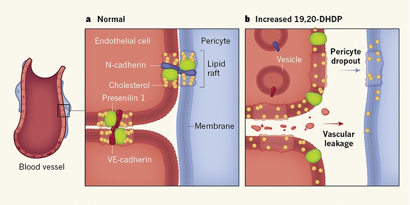
Major cause of dementia discovered

An international team of scientists have confirmed the discovery of a major cause of dementia, with important implications for possible treatment and diagnosis.

22 hours ago
 

'Man flu' may be real

The much-debated phenomenon of "man flu" may have some basis in fact, suggests an article published in the Christmas issue of The BMJ.

19 hours ago
popularity

Full moon linked to increased risk of fatal motorcycle crashes

The full moon is associated with an increased risk of fatal motorcycle crashes in the United States, the United Kingdom, Canada and Australia, finds a study in the Christmas issue of The BMJ.

19 hours ago
 

Drug increases speed, safety of treatment for multiple food allergies

In a randomized, controlled phase-2 clinical trial, an asthma medication increased the speed and safety of a protocol used to treat children for several food allergies at once, according to a study by researchers at the Stanford ...

19 hours ago
 

Social media trends can predict tipping points in vaccine scares

Analyzing trends on Twitter and Google can help predict vaccine scares that can lead to disease outbreaks, according to a study from the University of Waterloo.

23 hours ago
 

Team identifies DNA element that may cause rare movement disorder

A team of Massachusetts General Hospital (MGH) researchers has identified a specific genetic change that may be the cause of a rare but severe neurological disorder called X-linked dystonia parkinsonism (XDP). Occurring only ...

23 hours ago
Medical Xpress

Mechanisms of acetaminophen toxicity unraveled

NEWS

Mechanisms of acetaminophen toxicity unraveled

December 12, 2017 /EINPresswire.com/ -- In the U.S. and around the world, the most common cause of acute liver failure is acetaminophen (APAP) over dose. This commonly used pain reliever is safe and effective when used at recommended doses, but if taken at toxic levels, either by accident or purposefully, acetaminophen overdose lead to liver cell damage, acute liver failure, and death.

A significant public health problem, APAP hepatotoxicity accounts for about half of all acetaminophen-related acute liver failure cases and contributes to around 70,000 hospitalizations in the U.S. every year. APAP overdose is responsible for 46 percent of all cases of acute liver failure in the U.S.

In an effort to better understand the mechanisms of APAP toxicity, and to apply that increased understanding to provide better interventions than currently available, researchers at the University of Kansas Medical Center (UKMC) studied a broad range of cellular toxicity mechanisms that could lead to APAP-induced acute liver failure and liver cell (hepatocyte) death.

The authors note that decades of investigations into the mechanisms of APAP-induced liver injury “have provided significant insight into the role of APAP metabolism” into the cascade of events that can lead to liver injury. However, more insight is needed.

“Although acetaminophen is safe and effective when taken at therapeutic doses, the therapeutic window for treating acetaminophen overdoses is quite narrow and an APAP overdose is highly toxic to the liver.” said Dr. Hartmut Jaeschke of the Department of Pharmacology, Toxicology and Therapeutics at UKMC. “Our research is aimed at gaining a better understanding the mechanisms involved in APAP-induced liver injury so that more effective therapeutic interventions can be developed.”

According to Dr. Jaeschhke and co-researcher Dr. Anup Ramachandran, also of the UKMC Department of Pharmacology, Toxicology and Therapeutics, when consumed at therapeutic doses, the majority of APAP is excreted through the kidneys. However, after an overdose of APAP the body’s metabolic pathways are saturated and a variety of subsequent reactions lead the body form “APAP-protein adducts.” Adducts are products of a direct addition of two or more distinct molecules, resulting in a single “reaction product” containing all atoms of the components.

The initial oxidative stress caused by APAP-protein adducts occurs in the cells’ mitochondria, the site of cellular energy generation. According to the researchers, mitochondria can also play significant roles in cellular signaling and mitochondrial stress has emerged as a key factor in the cell signaling mechanism involved in APAP-induced liver cell death. This stress, however, has cellular ramifications, including mitochondrial failure.

“A better understanding of APAP-protein adduct formation and their relationship to liver cell death is very important,” explained Ramachandran. “Their formation on mitochondrial proteins is most relevant for understanding how APAP toxicity can lead to hepatocyte death.”

With APAP toxicity and mitochondrial APAP-protein adduct formation comes oxidative stress and the induction of “mitochondrial permeability transition pore” (MPTP), which ultimately compromises the mitochondrial membrane and shuts down mitochondrial function. Thus, MPTP is central to understanding APAP-induced liver injury, said the authors, who also note that it is now evident that the mitochondrial organelle also plays a significant role in the recovery and regeneration process after toxic injury.

On the positive side, the formation of APAP-protein adducts and their release into the circulatory system might be useful “biomarkers” for diagnosing APAP overdose. Too, they suggested that APAP overdose is a “clinically relevant model” for studying other causes of liver cell death and livery injury and is a model that can be used to study and test potential therapeutic intervention strategies.

What does their work mean for improving clinical interventions to save the lives of those with acute liver failure due to APAP toxicity?

“It is critical to connect these newly-discovered mediators and pathways of APAP metabolism to the already established mechanisms,” concluded the authors. “Although more can be learned about various aspects of these mechanisms, it is important to keep in mind potential effects of intervention strategies on drug metabolism, which can lead to misinterpretations. The relevance of these studies will, therefore, depend on the solid understanding of the various toxicity mechanisms.”


Their paper appears in the current issue of Gene Expression: The Journal of Liver Research. It is freely available on-line as an unedited, early epub at: http://www.ingentaconnect.com/content/cog/ge/

Antibiotics during pregnancy and risk of spontaneous abortion

Use of antibiotics during pregnancy and risk of spontaneous abortion

Flory T. Muanda, Odile Sheehy and Anick Berard

Severe Acne May Boost Prostate Cancer Risk

Severe Acne May Boost Prostate Cancer Risk

Renal & Urology News

Severe acne in adolescence may greatly increase an individual's risk of prostate cancer later in life, according to research published in the International Journal of Cancer.

The etiology of prostate cancer is poorly understood, though recent research suggests that Propionibacterium acnes, which is associated with acne vulgaris, is more likely to be present in the prostatic tissue of patients with the disease than in healthy individuals.

For this prospective population-based study, researchers reviewed the data of 284,198 males born between 1952 and 1956. Nearly all (more than 99% of) included individuals underwent conscription examinations in the 1970s; most individuals were examined at 18 or 19 years old. After excluding individuals with another primary malignancy prior to prostate cancer, missing data, or errors in health information, 243,187 were included in the analysis.


Reference

Nerves drive prostate cancer

 Nerves drive prostate cancer

Date:October 19, 2017
Source:Albert Einstein College of Medicine
                                                              
This image shows tissue from an early-stage tumor developing in a mouse model of prostate cancer. Sympathetic-nerve fibers (green) are closely intertwined with blood vessels (white). Norepinephrine released by nerve fibers stimulates vessel proliferation that fuels tumor growth.
In a study in today's issue of Science, researchers at Albert Einstein College of Medicine, part of Montefiore Medicine, report that certain nerves sustain prostate cancer growth by triggering a switch that causes tumor vessels to proliferate. Their earlier research -- which first implicated nerves in fueling prostate cancer -- has prompted Montefiore-Einstein to conduct a pilot study testing whether beta blockers (commonly used for treating hypertension) can kill cancer cells in tumors of men diagnosed with prostate cancer.

Journal Reference:

Ali H. Zahalka, Anna Arnal-Estapé, Maria Maryanovich, Fumio Nakahara, Cristian D. Cruz, Lydia W. S. Finley, Paul S. Frenette. Adrenergic nerves activate an angio-metabolic switch in prostate cancer. Science, 2017; 358 (6361): 321 DOI: 10.1126/science.aah5072
EDITED BY:M.HEZARKHANI   M.D. UROLOGIST

women with genetic risk, bi-annual MRI beats mammograms

NEWS

women with genetic risk, bi-annual MRI beats mammograms

December 8, 2017

Intensive surveillance including a dynamic contrast-enhanced magnetic resonance imaging (DCE-MRI) exam every six months was far more effective in detecting breast cancer in younger women with a high-risk genetic profile than an annual mammogram, according to a research team based at the University of Chicago Medicine and the University of Washington, Seattle.

University of Chicago Medical Cente

CUA guideline on the management of cystic renal lesions

Can Urol Assoc J. 2017 Mar-Apr; 11(3-4): E66–E73.
 2017 Mar 16. doi:  10.5489/cuaj.4484
PMCID: PMC5365391

CUA guideline on the management of cystic renal lesions


The Bosniak classification and management recommendations

Bosniak classification – key findingsRecommendations
Bosniak category I (simple renal cyst)  
  • Usually round or oval shape
  • Anechoic with posterior enhancement on US
  • Regular contour with clear interface with renal parenchyma
  • No septa, calcification or enhancement
  • No followup required

Bosniak category II  
  • Thin septum (
  • Fine calcification (often small, linear, parietal, or septal)
  • Small hypderdense cyst (20 HU)
  • No perceived contrast enhancement
  • No followup required

Bosniak category IIF  
  • Cyst unequivocally categorized as category II or III cysts
  • Multiple thin septa or a slightly thickened, but smooth septa
  • Calcifications – thick or nodular
  • No perceived contrast enhancement
  • Large hyperdense cysts (≥3 cm)
  • Followup recommended
  • Imaging at 6 months and 12 months after diagnosis and then annually for at least 5 years if no progression.

Bosniak category III  
  • Uniform wall thickening and/or nodularity
  • Irregular, thickened, and/or calcified septa
  • Contrast-enhancing sept
  • Surgical excision is suggested
  • Conservative management and RFA in select cases

Bosniak category IV  
  • Wall-thickening
  • Gross, irregular, and nodular septal thickening
  • Solid contrast-enhancing component, independent of septa
  • Malignant until proven otherwise
  • Surgical excision is suggested
  • Potential role for pretreatment RTB (of solid component) to confirm malignancy
  • RFA and conservative management in select cases

US: ultrasound; RFA: radiofrequency ablation; RTB: renal tumour biopsy.

EDITED BY:M.HEZARKHANI   M.D. UROLOGIST

Bosniak Classification for Complex Renal Cysts Reevaluated

J Urol. 2017 Jul;198(1):12-21. doi: 10.1016/j.juro.2016.09.160. Epub 2017 Mar 9.

Bosniak Classification for Complex Renal Cysts Reevaluated: A Systematic Review.


Abstract

PURPOSE:

We systematically evaluated the Bosniak classification system with malignancy rates of each Bosniak category, and assessed the effectiveness related to surgical treatment and oncologic outcome based on recurrence and/or metastasis.

MATERIALS AND METHODS:

In a systematic review according to PRISMA (Preferred Reporting Items for Systematic Reviews and Meta-Analyses) statement and the QUADAS-2 (Quality Assessment of Diagnostic Accuracy Studies) criteria, we selected 39 publications for inclusion in this analysis and categorized them into 1) surgical cohorts-all cysts treated surgically and 2) radiological cohorts-cysts with surgical treatment or radiological followup.

RESULTS:

A total of 3,036 complex renal cysts were categorized into Bosniak II, IIF, III and IV. In surgical and radiological cohorts pooled estimates showed a malignancy prevalence of 0.51 (0.44, 0.58) in Bosniak III and 0.89 (0.83, 0.92) in Bosniak IV cysts, respectively. Stable Bosniak IIF cysts showed a malignancy rate of less than 1% during radiological followup (surveillance). Bosniak IIF cysts, which showed reclassification to the Bosniak III/IV category during radiological followup (12%), showed malignancy in 85%, comparable to Bosniak IV cysts. The estimated surgical number needed to treat to avoid metastatic disease of Bosniak III and IV cysts was 140 and 40, respectively.

CONCLUSIONS:

The effectiveness of the Bosniak classification system for complex renal cysts was high in categories II, IIF and IV, but low in category III, and 49% of Bosniak III cysts was overtreated because of a benign outcome. This surgical overtreatment combined with the excellent outcome for Bosniak III cysts may suggest that surveillance is a rational alternative to surgery. This will require further study to assess whether surveillance of Bosniak III cysts will prove safe.

Copyright © 2017 American Urological Association Education and Research, Inc.

NEWS

Male-pattern baldness and premature graying associated with risk of early heart disease

Both were stronger risk factors than obesity

Date:November 30, 2017
Source:European Society of Cardiology
Summary:Male-pattern baldness and premature greying are associated with a more than fivefold risk of heart disease before the age of 40 years, according to new research. Obesity was associated with a fourfold risk of early heart disease

Binding of FUN14 Domain Containing 1 With Inositol 1,4,5-Trisphosphate Receptor in Mitochondria-Associated Endoplasmic Reticulum Membranes Maintains Mitochondrial Dynamics and Function in Hearts in Vivo


Mitochondrial protein in cardiac muscle cells linked to heart failure

Shengnan Wu, Qiulun Lu, Qilong Wang, Ye Ding, Zejun Ma, Xiaoxiang Mao, Kai Huang, Zhonglin Xie, Ming-Hui Zou

2017 American Heart Association, Inc

ارتباط افزایش خطر ابتلا به سرطان مثانه و آرسنیک موجود در آب چاه


طبق گزارش NCI ، نتایج تحقیقاتی که در New England انجام شده نشان می دهد نوشیدن آب چاه می تواند خطر ابتلا به سرطان مثانه را افزایش دهد. آب چاه ممکن است در سطوح کم یا متوسط حاوی آرسنیک باشد . ارتباط مصرف آب با سطح بالای آرسنیک با افزایش خطر ابتلا به سرطان مثانه اثبات شده است .
دو منبع احتمالی برای آرسنیک موجود در آب چاه وجود دارد. آرسنیک می تواند به طور طبیعی از سنگ های عمقی زمین ویا آفت کش های مبتنی بر آرسنیک که در مزارع سیب و سیب زمینی استفاده شود به درون آب نفوذ کند .
مداخلات موثری برای کاهش میزان آرسنیک در آب از طریق فیلتراسیون وجود دارد. با این حال ، استعمال دخانیات رایجترین و قویترین عامل خطر برای ابتلا به سرطان مثانه و ترک سیگار بهترین روش برای کاهش خطر سرطان مثانه است .

موسسه تحقیقات،درمان وآموزش سرطان

نقش پروتئین حمل کننده روی در مبارزه با سرطان پانکراس


عناصر کمیاب برای سلامتی بدن ضروری هستند. افزایش سطح عناصر کمیاب بدن و رسیدن به سطح سمی ، سلامتی را به خطر می اندازد. کنترل سطح این مواد می تواند اقدام موثری درمبارزه با بیماری ها باشد.
بیماران مبتلا به آلزایمر و پارکینسون نسبت به افراد سالم مقدار قابل توجهی آهن و روی در مغز خود دارند.
روی دومین عنصرکمیاب بعد از آهن است. افراد مبتلا به سرطان پانکراس ، مقدار زیادی از یک نوع خاص پروتئین حمل کننده روی را دارند. با کشف چگونگی حفظ این مواد در سطح مناسب و بررسی اثرات سطوح بالای این عناصر می توان مکانیسم حیاتی ZIP را رمزگشایی کرد . فرکانس رونویسی ZIP4 در سلولهای سرطانی پانکراس افزایش می یابد و جهش های ZIP4 منجر به اختلالات ژنتیکی کشنده می شود در حالیکه این اتفاق در بافت پانکراس طبیعی رخ نمی دهد .
ZIP4 یک هدف اصلی دارویی است که می تواند در درمان بسیاری بیماری ها کمک کند .

نتایج این مطالعه تحقیقاتی در نشریه Science Advances منتشر شده است .

September 6, 2017

موسسه تحقیقات،درمان وآموزش سرطان

The immune system and bone homeostasis

Rosebella A.IsemeabMarkMcevoybcBrianKellybdLindaAgneweFrederick R.WalkerbfgJohnAttiabch

aDepartment of Population and Reproductive Health, School of Public Health, Kenyatta University, P.O. Box 43844 –, 00100, Nairobi, Kenya
bSchool of Medicine & Public Health, The University of Newcastle, Callaghan, NSW, 2308, Australia
cHunter Medical Research Institute, Kookaburra Circuit, New Lambton Heights, NSW, 2305, Australia
dCentre for Brain and Mental Health Research, The University of Newcastle, Callaghan, NSW, Australia
eBrain Behaviour Research Group, School of Science and Technology, University of New England, Armidale, NSW 2351, Australia
fLaboratory of Affective Neuroscience, The University of Newcastle, Callaghan, NSW, Australia
gUniversity of Newcastle, Medical Sciences MS413, University Drive, Callaghan, NSW 2308, Australia
hDepartment of General Medicine, John Hunter Hospital, New Lambton Heights, NSW, Australia

 16 October 2017


Highlights

•Immune and bone systems recognised to share regulatory mechanisms and progenitor molecules.
•Osteoclasts and osteoblasts are continuously controlled by a variety of cells of the immune system.
•Altered bone composition often observed to occur alongside diseases characterised by immune dysfunction.
•Immune disturbance linked to pathogenic bone loss through the action of inflammatory markers and autoantibodies.


In a normal physiological state, the skeletal system carries out multiple functions including providing support, mobility, and protection for vital organs, as well as acting as a mineral reservoir for calcium and phosphate . In order to effectively carry out these tasks, the skeleton exists in a dynamic equilibrium characterised by continuous osteoclast-mediated bone resorption and osteoblast-mediated bone deposition. The latter biological process, termed “bone remodelling”, occurs in a harmonious and simultaneous fashion, resulting in a negligible change in bone mass. After peak bone mass is reached at the end of the 3rd decade, the normal balance between bone formation and bone resorption changes with relative increases in bone resorption leading to net bone loss . Interestingly, according to recent research, the majority of bone loss after the age of 65 is cortical bone loss; however, bone loss after menopause is mainly trabecular bone loss .

Briefly, bone remodelling follows the activation-resorption-formation (ARF) sequence . The first step, called the activation phase, begins with stimulation of quiescent osteoblasts. In response to appropriate stimuli, the latter release key osteoclast differentiation factors triggering preosteoclast fusion and differentiation to multinucleated osteoclasts marking the end of the activation phase . Once differentiated, osteoclasts polarize, adhere to the bone surface, and dissolve bone as part of the resorption phase. They then undergo apoptosis, as a means to prevent excessive bone resorption . After this resorptive process, there is an intermediate phase preceding bone formation, called a reversal phase. At this time, some macrophage-like uncharacterized mononuclear cells are observed at the site of remodelling, whose function consists of removal of debris produced during matrix degradation . The final phase termed bone formation is triggered by several growth factors stored in the bone matrix and released after its degradation, which are likely to be responsible for recruitment of osteoblasts in the resorbed area. Once recruited, osteoblasts produce new bone matrix, initially not mineralized (osteoid), and then they promote its mineralization, thus completing the bone remodelling process. Both cortical and trabecular bone undergo a continuous process of structural remodelling as a means of maintiaing mineral homeostasis, adapting to mechanical changes and repairing damage to bone.

Research has successfully highlighted various shared molecules and characteristics between bone and immune systems. Firstly, osteoclasts have been identified to derive from the same myeloid precursor cells that give rise to macrophages and myeloid dendritic cells . Moreover, osteoclasts are observed to exhibit the same lifecycles as dendritic cells, regulated by a variety of cytokines, transcription factors and inflammatory mediators. On the other hand, bone forming osteoblasts are noted to derive from mesenchymal stem cells.

The coordinated stimulation of osteoclasts by osteoblasts and reciprocal activation of osteoblasts by osteoclasts is referred to as coupling . It is widely acknowledged that the complementary regulatory activities of osteoclasts and osteoblasts are continuously controlled through direct cell to cell contact, via extracellular matrix interaction as well as by a variety of cells of the immune system . Osteoclasts represent the sole bone-resorbing cells in the body and are derived mainly following stimulation by two essential cytokines: the macrophage colony-stimulating factor (M-CSF) and the receptor activator of nuclear factor-kappa beta (RANK) ligand (RANKL), produced by osteoblasts. In particular, M-CSF aided by transcription factor PU.1 is responsible for driving the commitment of osteoclast progenitor cells . Whilst M-CSF stimulates proliferation of osteoclast precursors and upregulates RANK expression, PU.1 positively regulates the transcription of M-CSF receptor Colony stimulating factor 1 receptor, namely, c – Fms . Osteoporotic hormones such as 1,25 dihydroxyvitamin D3 (1,25(OH)2 D3), parathyroid hormone (PTH) and prostaglandin E2 (PGE2) are responsible for upregulating expression of RANKL.

Though the RANK/RANKL pathway represents the central process through which bone loss is regulated, there exist additional costimulatory pathways that are capable of modifying the net outcome resulting from bone remodelling. Osteoblast regulation of osteoclastogenesis additionally occurs through: interactions between immunoglobulin like receptors associated with immunoreceptor tyrosine based activation motif (ITAM) harbouring adaptor molecules (such as DNAX activating protein of 12KDa, known as DAP12 and Fc receptor common gamma subunit); via interactions between semaphorin 6D and its receptor plexin A1; and interactions between ephrin receptor B4 and ephrin B2 newly identified protein mediators of osteoblast – osteoclast interactions .

Additionally, a number of other cytokines are also reported to play a role in osteoclastogenesis. For example, tumor growth factor beta (TGFβ) is implicated in the enhanced recruitment of osteoblast progenitor cells to sits of bone resorption, whilst, tumor necrosis factor-α (TNF-α), is believed to facilitate the trafficking of osteoclast precursors from bone marrow to lymphoid organs. RANKL expression which plays a central role in osteoclast biology, has also been observed on various subsets of proliferative T cells (CD8 and CD4, Th (helper) 1 and Th2 cells) as well as Forkhead box p3 (Foxp3) expressing regulatory T (Treg) cells . Similarly, RANKL is also expressed on B lineage cells (i.e. B220 + cells that in the bone marrow represent multiple populations of early B cells precursors, immature B cells and mature B cells) . The latter observations suggest a pro-osteoclastogenic and bone resorption role for these immune cells.

Additionally, osteoblast differentiation is achieved by the coordinated activity of runt related transcription factor 2/core binding factor alpha 1 (RunX2/cbfa1) and distal-less homeobox 5 (Dlx5) which carry out integral transcriptional regulation for the conversion of bone marrow mesenchymal stem cells to osteoprogenitors cells . The latter occurring following commitment of MSc towards osteo/chrondo progenitor outcome driven by the Wingless (Wnt) pathway and a number of bone morphogenetic proteins (BMP) . Subsequently, a zinc finger containing transcription factor operating downstream of RunX2 termed osterix that is predominantly expressed in bone and cartilage provides additional support for the transition of osteoprogenitors to osteoblasts . A range of cytokines (i.e., IL-1, IL-6, IL-4, and TNF-α, TGF-β) are also noted to aid in regulating the differentiation and function of osteoblasts. Osteoblasts are therefore also influenced by immune cells, although the physiological and pathological significance and the molecular mechanisms are less well understood than in the case of osteoclasts. The precise roles attributed to the range of immune cells implicated in bone remodelling.

Efficacy of subcutaneous injection of platelet-rich plasma in alopecia: A clinical and histological pilot study on a rat model with a six-month long-term follow-up experience

 11 November 2017
  • DOI: 10.1111/jocd.12425  

To assess the potential of platelet-rich plasma (PRP) subcutaneous injection of to treat alopecia and to evaluate local toxicity.

Materials-Methods

Twelve Hairless rats were used. At D0, we performed systematic clinical examination and divided the rat back into four quadrants (Q). We initiated subcutaneous injection using either PRP in PRPQ+, platelet-poor plasma (PPP) in PPPQ+, physiological serum (PS) in PSQ+, or no treatment (Q4). At D7, D14, D21, and D28 but also second month (M2), M3, M4, M5, rats had exactly the same injection procedure. Follow-up with PRP efficacy and toxicity at D28 and M6 using clinical and histological evaluation was performed.

Results

Hair density was significantly improved at D28 and at M6 for PRPQ+ vs PSQ+ (respectively, P = .0156 and P = .0313), PPPQ+ (respectively, P = .042 and P = .046). Significant histological improvement was observed between D28 and M6, for PRPQ+ vs PPPQ+ and PSQ+ for vessels (respectively, P = .0160 and P = .021), collagen (respectively, P = .0036 and P = .032), and epithelium (respectively, P = .0138 and P = .022) with no local toxicity.

Conclusion

Our study suggests that subcutaneous PRP injections using controlled concentration of platelets and leukocytes improve hair growth.


Journal of Cosmetic Dermatology

A small-molecule inhibitor of TRPC5 ion channels suppresses progressive kidney disease in animal models
Science  08 Dec 2017:
Vol. 358, Issue 6368, pp. 1332-1336
DOI: 10.1126/science.aal4178
 
Progressive kidney diseases, whether caused by obesity, hypertension, diabetes, or rare genetic mutations, often have the same outcome: The cells responsible for filtering the blood are destroyed. Reporting today in Science, a team led by researchers from the Broad Institute of MIT and Harvard, Brigham and Women's Hospital, and Harvard Medical School describes a new approach to prevent death in these essential kidney cells. Studying multiple animal models of kidney disease, the team discovered a compound that can impede loss of the filtration cells and restore kidney function. The work, inspired by an investigation into a genetic form of the condition, has the potential to affect therapeutic research for millions of people suffering from progressive kidney diseases.
 
Progressive kidney diseases are often associated with scarring of the kidney’s filtration unit, a condition called focal segmental glomerulosclerosis (FSGS). This scarring is due to loss of podocytes, cells critical for glomerular filtration, and leads to proteinuria and kidney failure.
 
Inherited forms of FSGS are caused by Rac1-activating mutations, and Rac1 induces TRPC5 ion channel activity and cytoskeletal remodeling in podocytes. Whether TRPC5 activity mediates FSGS onset and progression is unknown.
 
The researchers identified a small molecule, AC1903, that specifically blocks TRPC5 channel activity in glomeruli of proteinuric rats. Chronic administration of AC1903 suppressed severe proteinuria and prevented podocyte loss in a transgenic rat model of FSGS. AC1903 also provided therapeutic benefit in a rat model of hypertensive proteinuric kidney disease. These data indicate that TRPC5 activity drives disease and that TRPC5 inhibitors may be valuable for the treatment of progressive kidney diseases.
 
 
Edited by: M.Hezarkhani  M.D. UROLOGIST
TEHRAN  UNIVERSITY

Male infertility

Medical causes

Problems with male fertility can be caused by a number of health issues and medical treatments. Some of these include:

  • Varicocele. A varicocele is a swelling of the veins that drain the testicle. It's the most common reversible cause of male infertility. Although the exact reason that varicoceles cause infertility is unknown, it may be related to abnormal testicular temperature regulation. Varicoceles result in reduced quality of the sperm.

    Treating the varicocele can improve sperm numbers and function, and may potentially improve outcomes when using assisted reproductive techniques such as in vitro fertilization.

  • Infection. Some infections can interfere with sperm production or sperm health or can cause scarring that blocks the passage of sperm. These include inflammation of the epididymis (epididymitis) or testicles (orchitis) and some sexually transmitted infections, including gonorrhea or HIV. Although some infections can result in permanent testicular damage, most often sperm can still be retrieved.
  • Ejaculation issues. Retrograde ejaculation occurs when semen enters the bladder during orgasm instead of emerging out the tip of the penis. Various health conditions can cause retrograde ejaculation, including diabetes, spinal injuries, medications, and surgery of the bladder, prostate or urethra.

    Some men with spinal cord injuries or certain diseases can't ejaculate semen, even though they still produce sperm. Often in these cases sperm can still be retrieved for use in assisted reproductive techniques.

  • Antibodies that attack sperm. Anti-sperm antibodies are immune system cells that mistakenly identify sperm as harmful invaders and attempt to eliminate them.
  • Tumors. Cancers and nonmalignant tumors can affect the male reproductive organs directly, through the glands that release hormones related to reproduction, such as the pituitary gland, or through unknown causes. In some cases, surgery, radiation or chemotherapy to treat tumors can affect male fertility.
  • Undescended testicles. In some males, during fetal development one or both testicles fail to descend from the abdomen into the sac that normally contains the testicles (scrotum). Decreased fertility is more likely in men who have had this condition.
  • Hormone imbalances. Infertility can result from disorders of the testicles themselves or an abnormality affecting other hormonal systems including the hypothalamus, pituitary, thyroid and adrenal glands. Low testosterone (male hypogonadism) and other hormonal problems have a number of possible underlying causes.
  • Defects of tubules that transport sperm. Many different tubes carry sperm. They can be blocked due to various causes, including inadvertent injury from surgery, prior infections, trauma or abnormal development, such as with cystic fibrosis or similar inherited conditions.

    Blockage can occur at any level, including within the testicle, in the tubes that drain the testicle, in the epididymis, in the vas deferens, near the ejaculatory ducts or in the urethra.

  • Chromosome defects. Inherited disorders such as Klinefelter's syndrome — in which a male is born with two X chromosomes and one Y chromosome (instead of one X and one Y) — cause abnormal development of the male reproductive organs. Other genetic syndromes associated with infertility include cystic fibrosis, Kallmann's syndrome and Kartagener's syndrome.
  • Problems with sexual intercourse. These can include trouble keeping or maintaining an erection sufficient for sex (erectile dysfunction), premature ejaculation, painful intercourse, anatomical abnormalities such as having a urethral opening beneath the penis (hypospadias), or psychological or relationship problems that interfere with sex.
  • Celiac disease. A digestive disorder caused by sensitivity to gluten, celiac disease can cause male infertility. Fertility may improve after adopting a gluten-free diet.
  • Certain medications. Testosterone replacement therapy, long-term anabolic steroid use, cancer medications (chemotherapy), certain antifungal medications, some ulcer drugs and certain other medications can impair sperm production and decrease male fertility.
  • Prior surgeries. Certain surgeries may prevent you from having sperm in your ejaculate, including vasectomy, inguinal hernia repairs, scrotal or testicular surgeries, prostate surgeries, and large abdominal surgeries performed for testicular and rectal cancers, among others. In most cases, surgery can be performed to either reverse these blockage or to retrieve sperm directly from the epididymis and testicles.

Environmental causes

Overexposure to certain environmental elements such as heat, toxins and chemicals can reduce sperm production or sperm function. Specific causes include:


  • Industrial chemicals. Extended exposure to benzenes, toluene, xylene, pesticides, herbicides, organic solvents, painting materials and lead may contribute to low sperm counts.
  • Heavy metal exposure. Exposure to lead or other heavy metals also may cause infertility.
  • Radiation or X-rays. Exposure to radiation can reduce sperm production, though it will often eventually return to normal. With high doses of radiation, sperm production can be permanently reduced.
  • Overheating the testicles. Elevated temperatures impair sperm production and function. Although studies are limited and are inconclusive, frequent use of saunas or hot tubs may temporarily impair your sperm count.

    Sitting for long periods, wearing tight clothing or working on a laptop computer for long stretches of time also may increase the temperature in your scrotum and may slightly reduce sperm production.

Health, lifestyle and other causes

Some other causes of male infertility include:

  • Illicit drug use. Anabolic steroids taken to stimulate muscle strength and growth can cause the testicles to shrink and sperm production to decrease. Use of cocaine or marijuana may temporarily reduce the number and quality of your sperm as well.
  • Alcohol use. Drinking alcohol can lower testosterone levels, cause erectile dysfunction and decrease sperm production. Liver disease caused by excessive drinking also may lead to fertility problems.
  • Tobacco smoking. Men who smoke may have a lower sperm count than do those who don't smoke. Secondhand smoke also may affect male fertility.
  • Emotional stress. Stress can interfere with certain hormones needed to produce sperm. Severe or prolonged emotional stress, including problems with fertility, can affect your sperm count.
  • Weight. Obesity can impair fertility in several ways, including directly impacting sperm themselves as well as by causing hormone changes that reduce male fertility.

1998-2017 Mayo Foundation for Medical Education and Research (MFMER)

Canola oil linked to worsened memory and learning ability in Alzheimer's

December 7, 2017

Canola oil is one of the most widely consumed vegetable oils in the world, yet surprisingly little is known about its effects on health. Now, a new study published online December 7 in the journal Scientific Reports by researchers at the Lewis Katz School of Medicine at Temple University (LKSOM) associates the consumption of canola oil in the diet with worsened memory, worsened learning ability and weight gain in mice which model Alzheimer's disease. The study is the first to suggest that canola oil is more harmful than healthful for the brain.

"Canola oil is appealing because it is less expensive than other vegetable oils, and it is advertised as being healthy," explained Domenico Praticò, MD, Professor in the Departments of Pharmacology and Microbiology and Director of the Alzheimer's Center at LKSOM, as well as senior investigator on the study.

Elisabetta Lauretti et al, Effect of canola oil consumption on memory, synapse and neuropathology in the triple transgenic mouse model of Alzheimer's disease, Scientific Reports (2017). DOI: 10.1038/s41598-017-17373-3

NEWS

Spinal tap needle type impacts the risk of complications

December 6, 2017

Spinal tap needle type impacts the risk of complications

Photo of the two needles used in lumbar puncture: Left: Conventional. Right: Atraumatic Credit: McMaster University

The study published today in The Lancet says using atraumatic needles rather than conventional traumatic needles for lumbar punctures is just as effective and results in a significant decrease in complications such as the headaches.

The type of needle used during a lumbar puncture makes a significant difference in the subsequent occurrence of headache, nerve irritation and hearing disturbance in patients, according to a study by Hamilton medical researchers.

 They found the pencil-point atraumatic with the better tip design has been available for about 70 years, but few physicians have been using it because they have not been aware of its benefits over the conventional bevelled traumatic needles.

There is a more than 50 per cent reduction in the occurrence of headaches with the atraumatic needles, and also more than a 50 per cent reduction in patient readmissions and return to emergency rooms for narcotics or blood patches.

A , commonly known as a spinal tap, is a regular medical procedure used to diagnose and to treat disease. Post-dural puncture headaches appear in about 35 per cent of patients, sometimes causing debilitating pain that can lead to a return to hospital for painkillers or more invasive treatment.

Edited by:M.Hezarkhani M.D. UROLOGIST

NEWS


Campylobacter jejuni

This scanning electron microscope image shows the characteristic spiral, or corkscrew, shape of C. jejuni cells and related structures

The Microbiome of Potentially Malignant Oral Leukoplakia Exhibits Enrichment for Fusobacterium, Leptotrichia, Campylobacter, and Rothia Species

Abdrazak Amer1, Sheila Galvin2, Claire M. Healy2 and Gary P. Moran1*
  • 1Division of Oral Biosciences, School of Dental Science, Trinity College Dublin, Dublin Dental University Hospital, Dublin, Ireland
  • 2Division of Oral and Maxillofacial Surgery, Oral Medicine and Oral Pathology, School of Dental Science, Trinity College Dublin, Dublin Dental University Hospital, Dublin, Ireland

Oral leukoplakia presents as a white patch on the oral mucosa and is recognized as having significant malignant potential. Although colonization of these patches with Candida albicans is common, little is known about the bacterial microbiota of these patches. In the current study we analyzed the microbiome of oral leukoplakia in 36 patients compared to healthy mucosal tissue from the same patients and healthy control subjects to determine if specific microbial enrichments could be identified early in the malignant process that could play a role in the progression of the disease. This was carried out by sequence analysis of the V1–V2 region of the bacterial 16S rRNA gene using the Illumina MiSeq. Oral leukoplakia exhibited increased abundance of Fusobacteria and reduced levels of Firmicutes (Metastats P < 0.01). Candida colonization was also more prevalent in leukoplakia patients relative to healthy controls (P = 0.025). Bacterial colonization patterns on oral leukoplakia were highly variable and five distinct bacterial clusters were discerned. These clusters exhibited co-occurrence of Fusobacterium, Leptotrichia, and Campylobacter species (Pearson P < 0.01), which is strikingly similar to the microbial co-occurrence patterns observed on colorectal cancers . Increased abundance of the acetaldehydogenic microorganism Rothia mucilaginosa was also apparent on oral leukoplakias from lingual sites (P 0.0012). Severe dysplasia was associated with elevated levels of Leptotrichia spp. and Campylobacter concisus (P < 0.05). Oral leukoplakia exhibits an altered microbiota that has similarities to the microbiome of colorectal cancer.

NEWS

Infertility linked to higher risk of death among women

November 1, 2017

Women with a history of infertility have a 10 percent increased risk of death compared to those without reported infertility struggles, according to results of a new study from researchers at the Perelman School of Medicine at the University of Pennsylvania. The study, which examined the association between infertility and mortality as well as specific causes of death, also showed that women with a history of infertility have a 20 percent increased risk of cancer-related mortality. The results of the study were presented this week at the annual American Society of Reproductive Medicine Scientific Congress & Expo in San Antonio, Texas.

Immunologic and endocrine functions of adipose tissue: implications for kidney disease

  • Nature Reviews Nephrology
  • doi:10.1038/nrneph.2017.157
  • Published online:04 December 2017


Excess adiposity can induce adverse sequelae in multiple cell types and organ systems. The transition from the lean to the obese state is characterized by fundamental cellular changes at the level of the adipocyte. These changes affect the local microenvironment within the respective adipose tissue but can also affect nonadipose systems. Adipocytes within fat pads respond to chronic nutrient excess through hyperplasia or hypertrophy, which can differentially affect interorgan crosstalk between various adipose depots and other organs. This crosstalk is dependent on the unique ability of the adipocyte to coordinate metabolic adjustments throughout the body and to integrate responses to maintain metabolic homeostasis. These actions occur through the release of free fatty acids and metabolites during times of energy need — a process that is altered in the obese state. In addition, adipocytes release a wide array of signalling molecules, such as sphingolipids, as well as inflammatory and hormonal factors (adipokines) that are critical for interorgan crosstalk. The interactions of adipose tissue with the kidney — referred to as the adipo–renal axis — are important for normal kidney function as well as the response of the kidney to injury. Here, we discuss the mechanistic basis of this interorgan crosstalk, which clearly has great therapeutic potential given the increasing rates of chronic kidney disease secondary to obesity and type 2 diabetes mellitus.


Key points

  • Adipocytes are metabolically active cells; they produce signalling lipids and metabolites and secrete protein factors (adipokines)

  • The relative levels of these lipids, proteins and metabolites change under different nutritional and pathological states, with adipocytes integrating information regarding the metabolic status quo at any given time and adjusting their cellular physiological state to maintain systemic homeostasis

  • Adipocyte-derived factors establish complex paracrine and endocrine signalling axes between local cell types in adipose tissue and other organs, including the kidney

  • A number of adipokines, including leptin and adiponectin, have well-established effects on kidney function; adiponectin might also be produced locally within the kidney and exert important metabolic functions in an autocrine fashion

  • As a source of pro-inflammatory cytokines, adipose tissue might exert important effects on the inflammatory state of the kidney

  • The renin–angiotensin system is present in adipose tissue and mediates inflammation in response to nutritional interventions; activation of this axis triggers profound signalling events in the kidney

Figure 1: Distinct features of adipocytes.

 Figure 1

Three types of adipocytes are generally found in rodents and humans. White adipocytes have few mitochondria and high lipid content; they store energy in the form of triglycerides. Brown adipocytes contain a high amount of mitochondria and mitochondrial brown fat uncoupling protein 1 (UCP1), which uncouples ATP generation and thus dissipates energy as heat. Beige adipocytes contain rather high mitochondrial activity and UCP1 and thus contribute to thermogenesis. Cold exposure or β-adrenergic (βAR) stimuli induce beige adipogenesis, either from de novo adipogenesis or transdifferentiation of white adipocytes, a biological process referred to as 'beiging'. In addition, beige adipocytes can revert back to white adipocytes under obesogenic conditions in a reverse process referred to as 'whitening'. CKD, chronic kidney disease.


Figure 2: Mechanisms of crosstalk between adipocytes and the kidney.


Figure 2

Adipose tissue exerts effects on the kidney through the actions of an array of secretory factors, such as cytokines, adipokines and metabolites — factors that at normal levels are important in maintaining kidney function. In response to excess caloric stress, adipose tissue expands through adipocyte hypertrophy and/or hyperplasia concomitantly with the development of insulin resistance and dysregulation of lipid metabolism. A shift in macrophage polarization from an anti-inflammatory M2 phenotype to a pro-inflammatory M1 phenotype also occurs, leading to the development of chronic inflammation through the excessive production of inflammatory cytokines and a reduction in anti-inflammatory adipokines, such as adiponectin. Adipocytes and macrophages coordinately orchestrate these inflammatory events and adipokine secretion. Renal dysfunction in the context of chronic kidney disease (CKD) is associated with high levels of adipose-derived molecules, such as leptin, adiponectin, angiotensin II, IL-6 and tumour necrosis factor (TNF) as well as dysregulated metabolites. Combined, these factors increase oxidative stress, inflammation and fibrotic transition in the kidney and eventually cause renal injury. Conversely, CKD exaggerates adipose tissue insulin resistance and inflammation potentially through the production of urea. Moreover, CKD promotes 'beiging' of adipose tissue, which favours energy loss and might therefore worsen kidney injury.


Figure 3: Angiotensin signalling in adiporenal crosstalk.

Figure 3


In addition to the liver, adipose tissue produces considerable amounts of angiotensinogen (AGT), which further converts to the active peptide angiotensin II (ANG II). Two types of ANG II receptors exist — type-1 angiotensin II receptors (AT1) and type-2 angiotensin II receptors (AT2) — which are present in both the kidney and adipose tissue and act antagonistically. Activation of AT1 signalling promotes macrophage infiltration in the kidney and adipose tissue and shifts polarization of M2 macrophages towards a pro-inflammatory M1 phenotype. Thus, ANG II–AT1 signalling accelerates inflammation and ultimately leads to glomerular injury and albuminuria. Moreover, alterations in adipose tissue function in the presence of ANG II, including adipose inflammation and hypertrophy, further exaggerate kidney injury. AT2 antagonizes AT1 signalling. In addition, AT1 signalling is blocked by type-1 angiotensin II receptor-associated protein (ATRAP), which increases internalization and desensitization of AT1.


Figure 4: Mechanisms of leptin function on kidney injury.

 Figure 4

Binding of leptin to its receptors (OBR; also known as LEP-R) in the kidney leads to activation of tyrosine-protein kinase JAK2 (JAK2) via phosphorylation, which activates signal transducer and activator of transcription 3 (STAT3) (and in some cases STAT5 (not shown)), which then translocates to the nucleus to regulate the transcription of target genes, including those encoding profibrotic factors. Activation of p38 mitogen-activated protein kinase (MAPK) via tyrosine-protein phosphatase non-receptor type 11 (SHP2; also known as PTPN11) and growth factor receptor-bound protein 2 (GRB2) might also promote STAT3 transcriptional activity. Phosphorylated JAK2 also activates NADPH oxidase and promotes oxidative signalling by generating reactive oxygen species (ROS). Independent of JAK2, leptin also activates the AKT–GSK3β pathway, which stabilizes β-catenin. Combined, these pathways increase the expression of transforming growth factor β-1 (TGFβ1) and other fibrogenic factors, such as collagen type IV (COL IV), fibronectin and vascular cell adhesion protein 1 (VCAM1). In a positive feedback loop, TGFβ1 binds to its receptor, TGFβR, and activates mothers against decapentaplegic homolog 2 (SMAD2) and SMAD3, further enhancing targeted gene expression. As a result, these dysregulated transcriptional programmes lead to inflammation and fibrogenesis in the kidney. High levels of circulating leptin lead to activation of renal mesangial cells and tubular inflammation. Moreover, leptin regulates kidney endothelial dysfunction, an important process that contributes to the development of kidney fibrosis. AKT, RAC-alpha serine/threonine-protein kinase; GSK3β, glycogen synthase kinase-3-β


Figure 5: Mechanisms of adiponectin actions in the kidney.


Figure 5

Adipocytes are the major source of circulating adiponectin, although the kidneys might also produce it. Adiponectin acts upon its receptors, ADIPOR1 and ADIPOR2, to activate 5′-AMP-activated protein kinase catalytic subunit α-1 (PRKAA1; also known as AMPK) and peroxisome proliferator-activated receptor-α (PPARα) signalling, respectively. Adiponectin receptors also possess ceramidase activity, which enables ceramides to be converted to sphingosines and fatty acids. Combined, these actions of adiponectin coordinately regulate energy metabolism, cellular oxidative stress and inflammation. Dysregulation of these cellular events can lead to various renal abnormalities, including fibrosis, glomerulosclerosis and albuminuria. Some compounds such as 5-aminoimidazole-4-carboxamide ribonucleotide (AICAR), resveratrol (RSV) and berberine (BBR) elevate circulating adiponectin levels and thus improve kidney dysfunction by enhancing adiponectin signalling. In addition, direct activation of AMPK by compounds such as PF-06409577 and PF-249 also ameliorate kidney disease. CaMKK 2, calcium/calmodulin-dependent protein kinase kinase 2; LBK1 (also known as STK11), serine/threonine-protein kinase STK11.

The expansion of adipose tissue is a major risk factor for an array of obesity-associated comorbidities, including type 2 diabetes mellitus (T2DM) and hypertension, that are risk factors for the development of chronic kidney disease (CKD). According to the World Health Organization, >1.9 billion adults aged ≥18 years were overweight (defined as a BMI ≥25 kg/m2) in 2014. Of these individuals, over 600 million were obese (BMI ≥30 kg/m2). The prevalence of obesity-related glomerulopathy increased greater than tenfold from 0.2% in 1986–1990 to 2.0% in 1996–2000 and to 2.7% in 2001–2015 . Moreover, >30% of patients undergoing kidney transplantation in the USA are obese with a BMI ≥35 kg/m2. Obesity and obesity-related glomerulopathy are in theory preventable. However, dozens of studies have highlighted the difficulties of losing weight and have demonstrated that the majority of individuals will ultimately regain lost weight even upon successful weight loss. Nevertheless, even transient weight loss induced by exercise, lifestyle modifications and dietary interventions significantly reduces proteinuria and glomerular hyperfiltration in obese individuals. A reduction of excess fat mass is therefore unquestionably beneficial in preventing pathological changes in the kidney through, for example, improvements in blood pressure and insulin sensitivity and through enhanced adipose tissue function associated with an improved adipokine profile.


EDITED BY:M.Hezarkhani    M.D. UROLOGIST

Environmental influences on ovarian dysgenesis — developmental windows sensitive to chemical exposures

  • Nature Reviews Endocrinology 13, 400414 (2017)
  • doi:10.1038/nrendo.2017.36


A woman's reproductive health and ability to have children directly affect numerous aspects of her life, from personal well-being and socioeconomic standing, to morbidity and lifespan. In turn, reproductive health depends on the development of correctly functioning ovaries, a process that starts early during fetal life. Early disruption to ovarian programming can have long-lasting consequences, potentially manifesting as disease much later in adulthood. A growing body of evidence suggests that exposure to chemicals early in life, including endocrine-disrupting chemicals, can cause a range of disorders later in life, such as those described in the ovarian dysgenesis syndrome hypothesis. In this Review, we discuss four specific time windows during which the ovary is particularly sensitive to disruption by exogenous insults: gonadal sex determination, meiotic division, follicle assembly and the first wave of follicle recruitment. To date, most evidence points towards the germ cell lineage being the most vulnerable to chemical exposure, particularly meiotic division and follicle assembly. Environmental chemicals and pharmaceuticals, such as bisphenols or mild analgesics (including paracetamol), can also affect the somatic cell lineages. This Review summarizes our current knowledge pertaining to environmental chemicals and pharmaceuticals, and their potential contributions to the development of ovarian dysgenesis syndrome. We also highlight knowledge gaps that need addressing to safeguard female reproductive health.


Key points

  • Humans are continuously exposed to numerous chemicals, including endocrine-disrupting chemicals (EDCs), throughout life

  • The ovarian dysgenesis syndrome hypothesis proposes that exposure to certain chemicals, including EDCs, during fetal life can lead to reproductive disorders later in life

  • Female fetuses are particularly sensitive to chemical exposure during four developmental windows: early gonadal development, meiotic division of germ cells, follicular assembly and early follicle recruitment

  • Some human epidemiological data suggest that the incidence of female reproductive disorders has increased since the mid-1950s and that EDCs are potential contributors

Figure 1

The ovarian dysgenesis syndrome hypothesis is based on the underpinning concept that early disruption of ovarian structure or function, caused by either genetic or environmental factors, leads to the impairment of reproductive function later in life. Alongside genetic mutations, several processes during development are sensitive to perturbation from chemical exposure. Such perturbation can result in disrupted migration or proliferation of primordial germ cells, disrupted gonadal sex differentiation, delayed or reduced follicle assembly, disturbed meiosis and accelerated folliculogenesis. PCOS, polycystic ovary syndrome; POI, premature ovarian insufficiency.

A dark side to omega-3 fatty acids

Nature.com

NEWS AND VIEWS

Diabetes can have many dangerous complications, including the progressive deterioration of blood vessels in the retina, which can lead to visual impairment. Despite the fact that this condition, called diabetic retinopathy, is a major cause of blindness in middle age, the pathways that mediate its development and progression are poorly understood. In a paper online in Nature, Hu et al. report that a derivative of the omega-3 fatty acid docosahexaenoic acid (DHA) has a role in diabetic retinopathy.

Dietary intake of DHA is associated with several health benefits, including normal brain and eye function. Moreover, mice that lack the protein Mfsd2a, which transports DHA across the blood–brain barrier into the brain, develop severe retinal and brain dysfunction. Omega-3 fatty acids such as DHA have key functions in cell membranes, where they provide membrane fluidity and flexibility, and promote the membranes’ role as barriers, allowing the selective transport of molecules into and out of cells.

In addition, omega-3 fatty acids can have signalling roles. DHA is highly susceptible to oxidation, producing ‘lipid mediator’ molecules that can regulate cell–cell signalling — for instance, to orchestrate the return to normal conditions following an inflammatory reaction. One class of lipid mediator produced by DHA oxidation is epoxide molecules. These metabolites are rapidly degraded by the enzyme soluble epoxide hydrolase (sEH) to form dihydroxy derivatives such as 19,20-dihydroxydocosapentaenoic acid (19,20-DHDP). Indeed, 19,20-DHDP is the major product of DHA metabolism in the retina.

Given that DHA and epoxide metabolites are associated with beneficial retinal effects, might sEH play a part in diabetic retinopathy by reducing the levels of these molecules? Hu et al. set out to examine this possibility using a mouse model of the disease. They found that increases in sEH expression in Müller glia cells (which support neuronal function in the retina) correlated with the onset of several early signs of diabetic retinopathy. Such changes included: the degeneration of pericyte cells, which wrap around and provide support for the endothelial cells that make up the blood-vessel wall; an increase in the number of acellular capillaries, which have abnormal transport and barrier properties; and leakage of plasma out of vessels into the surrounding retinal tissue. These defects eventually lead to poor circulation in the retina, abnormal and uncontrolled blood-vessel formation, retinal bleeding and visual impairment. 

Importantly, the authors demonstrated that pharmacological inhibition of sEH or genetic suppression of the gene encoding sEH prevented these pathological events, suggesting that sEH has a causal role in diabetic retinopathy. How might it exert these effects? Hu and colleagues showed that it is the production of 19,20-DHDP, rather than the reduction in DHA or epoxide molecules, that is the key trigger of vascular degeneration. 

Next, the group found that the mechanism of pathogenesis involves interactions between three proteins — presenilin 1, vascular endothelial (VE)-cadherin and neural (N)-cadherin. The two cadherins are an essential part of protein complexes called adherens junctions, which anchor cell–cell adhesion sites on cell surfaces to the cell interior. VE-cadherin is found at junctions between pairs of endothelial cells, and N-cadherin is located at junctions between endothelial cells and pericytes.

The authors showed that presenilin 1 and the cadherins associated with cholesterol in clusters called lipid rafts in cell membranes (Fig. 1a).

Figure 1 | Abnormal lipid dynamics in diabetic retinopathy. Hu et al.1 have shown that increases in levels of the molecule 19,20-DHDP promote the development and progression of a form of visual impairment called diabetic retinopathy in mice. a, Under normal conditions, the endothelial cells that line blood vessels in the retina are connected to one another and to supporting cells called pericytes through adhesion molecules known as cadherins in cell membranes. These connections rely on associations between the cadherins and the protein presenilin 1, which are, in turn, facilitated by clustering of cholesterol on ‘lipid rafts’. b, The authors show that, during the early stages of diabetic retinopathy, increases in 19,20-DHDP in cell membranes (not shown) alter the distribution of presenilin 1, perhaps by disrupting the distribution of cholesterol molecules. Presenilin 1, no longer restrained to lipid rafts, does not associate with cadherins, leading to cadherin internalization in vesicles. This destabilizes cell–cell contacts, resulting in pericyte ‘dropout’ from around vessels, and vascular leakage.

However, increased levels of 19,20-DHDP altered the biophysical properties of membranes, leading to decreased interactions between presenilin 1 and cadherins in lipid rafts, presumably by changing the distribution of cholesterol(Fig. 1b). This, in turn, led to internalization of cadherins into the cell, disrupting adherens junctions. Ultimately, this process weakened cell–cell adhesion, causing pericyte ‘dropout’ from the blood vessel and breaching of the vascular barrier.

Together, Hu and colleagues’ results indicate that 19,20-DHDP acts through a different mechanism from that of another player in retinopathy — vascular endothelial growth factor (VEGF).

This protein is induced under low-oxygen conditions and acts through receptors on vascular endothelial cells to induce proliferation, resulting in fragile new blood vessels prone to bleeding. Furthermore, the authors demonstrated that overexpression of sEH in the retinas of wild-type mice led to increased levels of 19,20-DHDP, vascular leakage and pericyte dropout, suggesting that sEH activation alone is sufficient to induce retinal-vessel defects, without VEGF-mediated mechanisms. Inhibiting VEGF halts the progression of diabetic retinopathy and that of another vision disorder caused by changes in retinal blood vessels: wet age-related macular degeneration. But not all patients respond to anti-VEGF therapy, and those who do often become insensitive to treatment over time, making Hu and colleagues’ discovery of an alternative pathway valuable.

Previous work has shown that a different lipid mediator that acts on endothelial cells, the molecule sphingosine 1-phosphate, induces the formation of adherens junctions and promotes the development of blood vessels. Hu and colleagues’ newly discovered role for omega-3 derivatives thus adds to the list of crucial lipid signalling pathways known to be involved in retinal vascular diseases. It is possible that both pathways could be targeted therapeutically. 

Hu and co-workers’ study raises interesting avenues for future research. First, what triggers increases in sEH production in diabetic retinopathy? Dysregulated metabolic control of blood glucose can cause oxidative stress (an inability to deal with free radicals produced during metabolism) in the retina, which might be a key factor. However, a molecular mechanism by which this could lead to increased sEH remains to be elucidated. Second, it is unclear how 19,20-DHDP is transported from Müller glia cells to endothelial cells or pericytes. The apparent specificity of 19,20-DHDP among all omega-3 fatty acid derivatives as a mediator of diabetic retinopathy is also not understood at the molecular level. Perhaps 19,20-DHDP relies on specific targets, such as particular cell-surface or nuclear receptors, for its functions. Such targets could be a part of separate pathways to the membrane-lipid effects uncovered by the authors 

In summary, Hu and colleagues’ findings suggest that the metabolism of DHA to 19,20-DHDP by sEH is one of the key regulatory systems that disrupt endothelial junctions at the blood–retinal barrier. The authors’ work might help to guide the development of therapeutic approaches that block the activity of sEH to control diabetic retinopathy and wet age-related macular degeneration. The study might even point to ways of treating neurological disorders, including stroke and neurodegenerative diseases, that are characterized by a compromised blood–brain barrier — similar mechanisms might be expected to operate in these diseases.


Edited by:M.Hezarkhani  M.D.  UROLOGIST


Metabolic syndrome and smoking are associated with an increased risk of nocturia in male patients with benign prostatic enlargement

  • Prostate Cancer and Prostatic Diseases (2017)
  • doi:10.1038/s41391-017-0003-z
Published online: 04 December 2017

To evaluate the relationship between cigarette smoking, metabolic syndrome (MetS) and nocturia in patients with lower urinary tract symptoms (LUTS) and benign prostatic enlargement (BPE).

Overall 492 patients were enrolled with median age and BMI of 68 years (IQR 61/74) and 26.5 kg/m2 (IQR: 24/29), respectively. Moderate/severe nocturia was reported in 212 (43.1%) patients. MetS was diagnosed in 147 (29.9%) patients and out of them 89 (60.5%) complained moderate/severe nocturia (p = 0.001). Overall 187 (38%) patients were current smokers and out of them 99 (52%) presented moderate/severe nocturia(p = 0.034). Patients with moderate/severe nocturia were older (p = 0.001) and with larger prostate volume (p = 0.003). On multivariate analysis, age (OR: 1.067 per year, 95% CI: 1.036–1.098; p = 0.001), PV (OR: 1.011 per ml, 95% CI: 1.003–1.019; p = 0.006), MetS (OR: 2.509, 95% CI: 1.571–4.007; p = 0.001) and smoking (OR: 1.690, 95% CI: 1.061–2.693; p = 0.027) were associated with nocturia severity.

MetS and smoking doubled the risk of moderate/severe nocturia in patients with LUTS and BPE. Assessing smoking and metabolic status in LUTS/ BPE patients is suggested.


Edited. by: M.Hezarkhani  M.D. Urologist

Nature.com/prostate cancer and prostatic diseases


Antibiotics administered during labor delay healthy gut bacteria in babies

Source: McMaster University

November 28, 2017

Early life microbial colonization and succession is critically important to healthy development, with impacts on metabolic and immunologic processes throughout life.

One out of every three or four pregnant women test positive for Group B Streptococcus during routine screening and the majority choose to receive antibiotic prophylaxis during labour to prevent GBS transmission to their infant at birth. Infant infections can lead to serious illness including meningitis and death in a very small number of infants, and antibiotic treatment is an important prevention strategy.

Our research indicates there is a delay in the expansion of the dominant infant gut colonizer, called Bifidobacterium, when infants are exposed to antibiotics for GBS prevention during vaginal labour.

It's a good sign that bacterial groups recover by 12 weeks but it's still unclear what these findings mean for infant health, especially since early infancy is such an important developmental time.

The study utilized data from 74 mother-infant pairs in the McMaster pilot cohort called Baby & Mi. Participants came from low-risk populations in Hamilton and Burlington, Ontario.

The gut bacteria development of the infants was tested at four points over the first 12 weeks of life, including at three days, 10 days, six weeks and 12 weeks.

The babies were healthy, full-term, breast-fed babies predominantly born vaginally, with a small percentage born by C-section that were also exposed to antibiotics to prevent surgical infection. As in previous studies, babies born by C-section had delayed expansion of the key gut colonizer compared to babies born vaginally without exposure.

A larger study is underway that will determine the long-term consequences of antibiotics administered during labour for GBS on both microbial succession and on health and disease risk. This will help us explore in greater depth the influence of maternal and infant variables on the infant gut microbiome.

Edited by:M.Hezarkhani  M.D. Urologist

Journal Reference:

Jennifer C. Stearns, Julia Simioni, Elizabeth Gunn, Helen McDonald, Alison C. Holloway, Lehana Thabane, Andrea Mousseau, Jonathan D. Schertzer, Elyanne M. Ratcliffe, Laura Rossi, Michael G. Surette, Katherine M. Morrison, Eileen K. Hutton. Intrapartum antibiotics for GBS prophylaxis alter colonization patterns in the early infant gut microbiome of low risk infants. Scientific Reports, 2017; 7 (1) DOI: 10.1038/s41598-017-16606-9

Stress during pregnancy affects the size of the baby

Source:University of New Mexico

November 28, 2017

prenatal maternal stress late in gestation causes mothers to invest less energy in their offspring, which leads to slower grow in the womb and during infancy. Once the baby has reached nutritional independence, however, they are no longer affected directly by their mother's provisioning, and consequently grow at the same rate as non-disadvantaged offspring. Thus, maternal stress late in gestation leads to slow growth during dependent phases, but doesn't affect growth later.

By contrast, prenatal maternal stress early in gestation additionally causes the fetus to be entirely reprogrammed to deal with a reduced life expectancy. To "make the best of a bad job," the early challenged offspring switches to an accelerated pace of life and grows and matures faster than unchallenged offspring to ensure that it reproduces before it dies. Once set on the fast track, the offspring under early prenatal maternal stress remain on this trajectory even after weaning and therefore overshoot the usual body size for age throughout development.

"These new results may bear some translational value for understanding why girls start their menstrual cycles earlier in poorer neighborhoods." In combination, an infant's acceleration of their developmental processes together with a deceleration due to reduced maternal investment could then cancel each other out during phases of intense maternal investment -- gestation and lactation. It is not until the infant is nutritionally independent that the programming effects become clear.

This new comparative study finds all of these predictions are supported in a large sample of studies that each measured the effects of prenatal stress on offspring size and growth compared to an unchallenged control group.

"We found that stress during late gestation reduces offspring growth during dependence, resulting in a reduced body size throughout development, whereas stress during early gestation results in largely unaffected growth rates during dependence but accelerated growth and increased size after weaning," says Berghänel.

All stressors seem to have the same effect, and the results are stable across a variety of experiments. Whether mothers were exposed directly to stressors via food restriction or other adversities or were experimentally manipulated to increase their "stress hormones" for example, cortisol, the patterns of offspring growth across developmental stage relative to the timing of the stressor remained the same.

These new results may bear some translational value for understanding why girls start their menstrual cycles earlier in poorer neighborhoods, why teenage pregnancies are more frequent in disadvantaged families, and why adverse conditions during early development, particularly in formula-fed children, often lead to obesity and other metabolic health problems later in life.

Maternal stress during gestation causes numerous effects on infant physiology that extend well into adulthood. Empirical tests of this hypothesis across mammals suggest that the timing of the stressor during gestation and a simultaneous consideration of maternal investment and adaptive growth plasticity effects are crucial for a full comprehension of prenatal stress effects on offspring growth. The results support an adaptive life history perspective on maternal effects that is relevant for evolutionary biology, medicine, and psychology.


Journal Reference:

Andreas Berghänel, Michael Heistermann, Oliver Schülke, Julia Ostner. Prenatal stress accelerates offspring growth to compensate for reduced maternal investment across mammals. Proceedings of the National Academy of Sciences, 2017; 201707152 DOI: 10.1073/pnas.1707152114

Edited by:M.Hezarkhani  M.D.  Urologist

Sperm RNA may serve as biomarkers of future health

Source:Wayne State University
December 1, 2017


At fertilization, sperm delivers a structurally distinct genome, along with a complement of ribonucleic acids, or RNAs, and proteins to the immature egg cell.

To test the hypothesis, sperm RNA elements corresponding to specific genes were characterized as a function of disease association. Dr. Krawetz's team surveyed a total of 278,605 sperm RNA elements called short exon-sized sequences, or SREs, associated with diseases. This functional association of SREs may indicate a future phenotype, providing improved understanding of the father's contribution to the life course of the child as well as the current state of paternal health.

In the future, if those SREs that are mutated or modified can be identified, researchers and physicians may be able to not only forecast disease or conditions, but develop ways to prevent them.

Edited by:M.Hezarkhani  M.D. Urologist

Blood flow altered in brains of preterm newborns vs. full-term infants

Blood, oxygen and nutrients follow function, with more flowing to rapidly developing cerebral regions

Date:
December 4, 2017
Source:
Children's National Health System
Summary:
Cerebral blood flow of key regions of newborns' brains is altered in very premature infants and may provide an early warning sign of disturbed brain maturation well before such injury is visible on conventional imaging, according to a prospective, observational study.
2017 ScienceDaily

Journal Reference:

  1. Marine Bouyssi-Kobar, Jonathan Murnick, Marie Brossard-Racine, Taeun Chang, Eman Mahdi, Marni Jacobs, Catherine Limperopoulos. Infants Born Preterm Compared with Infants Born Full Term. The Journal of Pediatrics, 2017 DOI: 10.1016/j.jpeds.2017.09.083

Lithium chloride blunts brain damage linked to fetal alcohol syndrome

Date:
December 5, 2017
Source:
NYU Langone Health / NYU School of Medicine
Summary:
A single dose of lithium chloride, a drug used to treat bipolar disease and aggression, blocks the sleep disturbances, memory loss, and learning problems tied to fetal alcohol syndrome, new experiments in mice show.
Published in the journal Neuroscience online Nov. 26
2017 ScienceDaily

G protein-coupled estrogen receptor regulates embryonic heart rate in zebrafish

 Published: October 24, 2017
 

Estrogens act by binding to estrogen receptors alpha and beta (ERα, ERβ), ligand-dependent transcription factors that play crucial roles in sex differentiation, tumor growth and cardiovascular physiology. Estrogens also activate the G protein-coupled estrogen receptor (GPER), however the function of GPER in vivo is less well understood. Here we find that GPER is required for normal heart rate in zebrafish embryos. Acute exposure to estrogens increased heart rate in wildtype and in ERα and ERβ mutant embryos but not in GPER mutants. GPER mutant embryos exhibited reduced basal heart rate, while heart rate was normal in ERα and ERβ mutants. We detected gper transcript in discrete regions of the brain and pituitary but not in the heart, suggesting that GPER acts centrally to regulate heart rate. In the pituitary, we observed gper expression in cells that regulate levels of thyroid hormone triiodothyronine (T3), a hormone known to increase heart rate. Compared to wild type, GPER mutants had reduced levels of T3 and estrogens, suggesting pituitary abnormalities. Exposure to exogenous T3, but not estradiol, rescued the reduced heart rate phenotype in gper mutant embryos, demonstrating that T3 acts downstream of GPER to regulate heart rate. Using genetic and mass spectrometry approaches, we find that GPER regulates maternal estrogen levels, which are required for normal embryonic heart rate. Our results demonstrate that estradiol plays a previously unappreciated role in the acute modulation of heart rate during zebrafish embryonic development and suggest that GPER regulates embryonic heart rate by altering maternal estrogen levels and embryonic T3 levels.


 summary

Estrogen hormones are important for the formation and function of the nervous, reproductive and cardiovascular systems. Here we report that acute exposure to estrogens increases heart rate, a previously unappreciated function of estrogens. Using zebrafish with mutations in genes that respond to estrogens, we found that heart rate is regulated not by the typical molecules that respond to estrogens–the nuclear estrogen receptors–but rather by a different molecule, the G protein-coupled estrogen receptor. We also show that estrogens increase heart rate by increasing levels of thyroid hormone.

Our results reveal a new function for the G protein-coupled estrogen receptor and a new connection between estrogens and thyroid hormone. Environmental compounds that mimic estrogens can be harmful because they can influence gonad function. Our results suggest that endocrine disrupting compounds may also influence cardiac function.

PLOS.ORG

Edited by:M.Hezarkhani  M.D.  Urologist